Description
Overview & Strengths
-
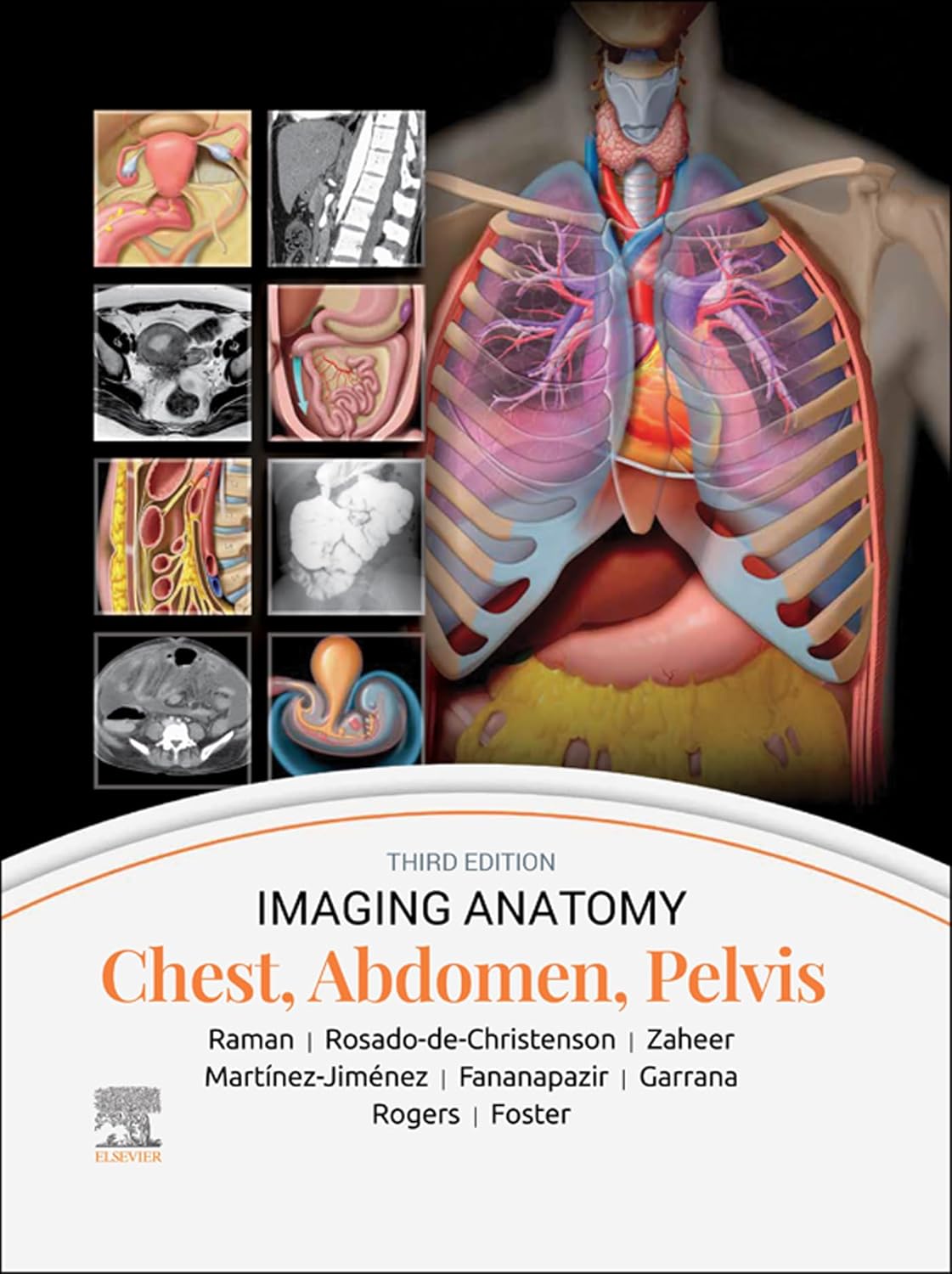
The atlas is “richly illustrated and superbly organized,” designed as an “excellent point-of-care resource for practitioners at all levels of experience and training”, offering fast access to anatomical reference in busy clinical environments.
-
Aimed at clarifying normal anatomy and anatomical variants, it provides detailed imagery across a variety of modalities—CT, MRI, ultrasound, 3D reconstructions, and medical illustrations—to help clinicians quickly identify and interpret findings.
-
The layout is meticulous and user-friendly, employing templated pages, concise bulleted text, and a highly formatted design that makes navigating complex anatomy fast and intuitive.
-
This edition includes nearly 2,800 images, and introduces updated content covering developmental anatomy, vascular variants, the PI-RADS system for prostate MRI, chest wall musculature, body MR applications, GU/GYN imaging nuances, and representative pathological examples.
📌 Key Features
-
2,800+ High-Quality Images – Includes CT, MRI, ultrasound, 3D reconstructions, and detailed medical illustrations.
-
Multimodality Coverage – Shows anatomy in multiple imaging techniques for better cross-referencing.
-
Clear Labels & Annotations – Enhances learning and quick identification of anatomical structures.
-
Templated Layout – Consistent format with bulleted text for rapid lookup in clinical settings.
-
Updated Content – New chapters and revisions on vascular variants, PI-RADS prostate MRI, GU/GYN imaging, and developmental anatomy.
-
Pathology Examples – Integrates selected pathological cases alongside normal anatomy for clinical context.
-
Anatomical Variants Highlighted – Prepares readers for recognizing normal variations that could mimic pathology.
-
Expert Authorship – Written by experienced radiologists with academic and clinical expertise.
-
Cross-Sectional Orientation Guides – Helps in correlating imaging planes with real anatomical positioning.
-
Digital Access (where available) – Some editions offer eBook versions for portable study.
🎯 Benefits of Reading
-
Boosts Diagnostic Accuracy – Improves ability to recognize normal vs. abnormal findings quickly.
-
Saves Time in Practice – Rapid reference format suits busy radiology and clinical environments.
-
Enhances Exam Prep – Perfect for residents and fellows studying for board or certification exams.
-
Strengthens Multidisciplinary Collaboration – Helps non-radiologist clinicians understand imaging reports more clearly.
-
Supports Teaching & Presentations – High-resolution images are useful for academic lectures.
-
Reduces Errors – Awareness of anatomical variants decreases misinterpretation risk.
-
Keeps Knowledge Current – Updated content reflects modern imaging standards and protocols.
-
Improves Procedural Planning – Essential for surgeons and interventionalists mapping anatomy before operations.
-
Facilitates Lifelong Learning – Comprehensive yet concise for both newcomers and experienced practitioners.
-
Increases Confidence – Knowing anatomy precisely in multiple imaging modalities builds clinical certainty.

Reviews
There are no reviews yet.